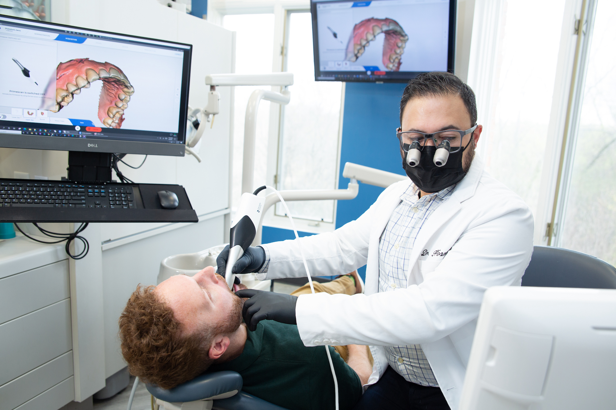
Planning oral surgery used to mean working from flat images and educated guesses. Not anymore. 3D imaging gives your care team a true-to-life view of your anatomy before a single incision is made. At Ariata Dental in Gurnee, Dr. Arash Hosseini uses 3D dental imaging to map bone, nerves, and sinuses down to the millimeter, so wisdom tooth removal and implant placement are safer, faster, and far more predictable.
What Is 3D Dental Imaging (CBCT)?
3D dental imaging, also called cone beam computed tomography (CBCT), is a scan that produces a complete three-dimensional view of your teeth, jawbone, nerves, sinuses, and airway in a single pass. Unlike flat 2D X-rays, which stack overlapping structures onto one image, CBCT captures depth and angulation, giving Dr. Hosseini a true picture of what’s happening beneath the surface.
The scan itself is quick and non-invasive. You sit or stand still while the scanner rotates once around your head, usually in under 30 seconds. Radiation exposure is significantly lower than a medical CT scan, making it a safe choice for most families and children, including teens being evaluated for wisdom teeth.
At Ariata Dental in Gurnee, we use 3D imaging for surgical planning, implant placement, airway evaluation, and diagnosing issues that simply don’t show up on traditional X-rays. It’s one of the tools that makes modern oral surgery so much more precise than the methods of even a decade ago.
How a 3D Scan Works: From Capture to Surgical Plan
A CBCT scan turns a short, still moment into a detailed surgical plan. Here’s how the process unfolds at Ariata Dental:
- Positioning and capture. You’ll be positioned in the scanner with your head gently stabilized. The cone-shaped beam rotates around your head, capturing hundreds of images from multiple angles in a single rotation.
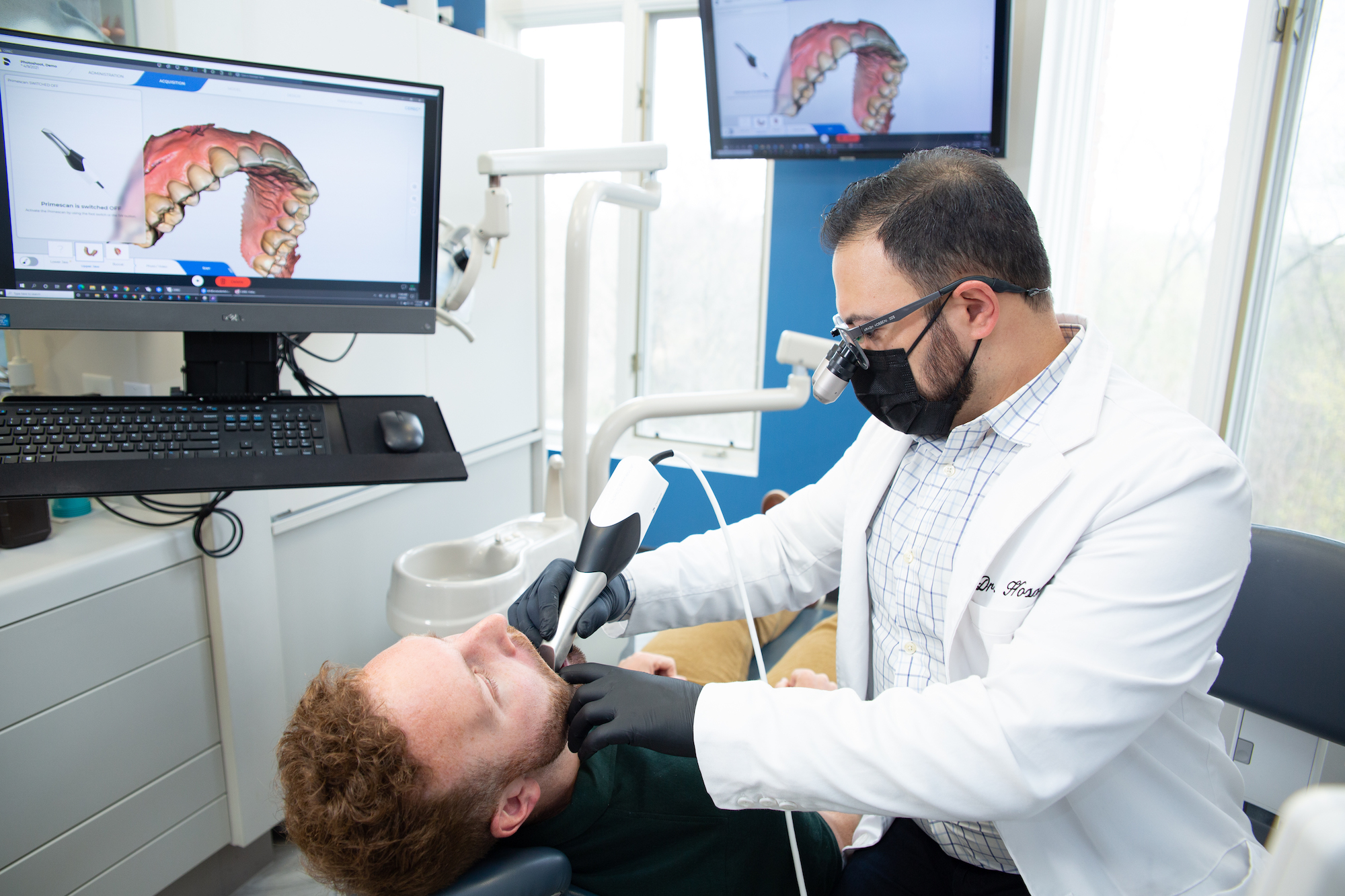
- 3D reconstruction. Specialized software stitches those images together into a true three-dimensional model of your teeth, jawbone, nerves, and sinuses. Dr. Hosseini can rotate, slice, and zoom into any part of the model.
- Measurement and analysis. Using the scan, Dr. Hosseini measures bone density, the exact distance to the inferior alveolar nerve, sinus floor height, and the angulation of impacted teeth. These numbers directly shape the surgical approach.
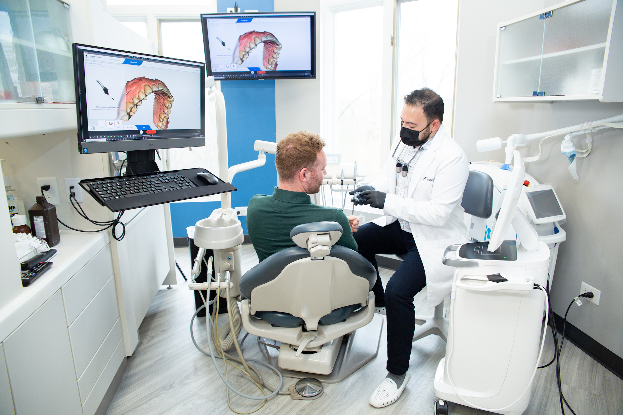
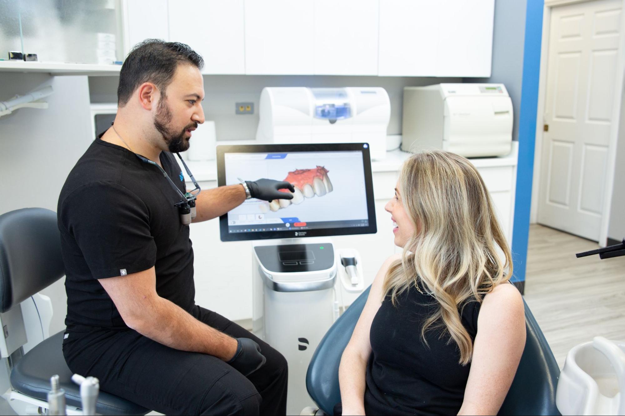
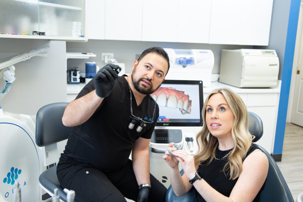
- Patient review. You’ll see the 3D model during your consultation. Dr. Hosseini walks you through what he sees, what the plan is, and why. It’s much easier to understand your own anatomy when you can see it from every angle.
- Long-term record. Your scan is stored securely for future comparison, follow-up visits, and ongoing treatment planning.
Key Benefits of 3D Imaging for Oral Surgery
The advantages of CBCT go well beyond nice pictures on a screen. 3D imaging can translate directly into better outcomes for you. The benefits touch nearly every stage of your care, from diagnosis through recovery, and they make a real difference in how comfortable and confident you feel going into surgery.
- Higher diagnostic accuracy. Impactions, cysts, hidden infections, bone loss, and anatomical variations that are invisible on 2D X-rays become obvious in 3D. That means fewer surprises during surgery.
- Precise nerve and sinus mapping. The inferior alveolar nerve runs near the lower wisdom teeth, and the sinus floor sits just above upper molars. Knowing exactly where these structures are reduces the risk of nerve injury or sinus perforation.
- Guided implant placement. When implants are placed using 3D planning and a custom guide, they sit in the ideal position for strength, aesthetics, and long-term success. This is one of the biggest advances in implant dentistry.
- Smaller, less invasive incisions. Because Dr. Hosseini knows exactly where to go, surgery can often be performed through smaller openings with less tissue disruption.
- Shorter chair time. Pre-planning means less time spent making decisions during surgery. You’re in and out faster.
- Faster recovery and less post-op discomfort. A less invasive technique typically leads to less swelling, less bruising, and a smoother healing experience.
Beyond the clinical advantages, two more benefits are worth calling out, because they shape how you feel about the whole experience. Better communication during your consultation means you leave informed instead of confused. Seeing your own 3D scan makes it easier to understand what’s happening and what the plan is. And for complex cases, like patients with prior failed implants, unusual anatomy, or dense bone, 3D imaging makes the difference between guessing and knowing. That’s the kind of confidence you want on both sides of the treatment chair.
3D Imaging vs. Traditional 2D X-rays
Both 2D X-rays and CBCT scans have a place in modern dentistry. The short answer: 2D X-rays work well for everyday checkups and spotting decay, while 3D imaging is essential for surgical planning and anything involving bone, nerves, or implants. Here’s how they compare side by side.
| Feature | 2D X-rays (Panoramic/Bitewing) | 3D CBCT Scan |
|---|---|---|
| View type | Flat, overlapping image | True three-dimensional model |
| Detail level | Good for decay and everyday anatomy | Reveals nerves, sinuses, bone density, airway |
| Radiation dose | Very low | Higher than 2D, much lower than medical CT |
| Scan time | Seconds | Under 30 seconds |
| Best use case | Routine exams, cavity checks | Implants, wisdom teeth, complex surgery |
For a routine cleaning and exam, a 2D X-ray is the right tool. For wisdom tooth evaluation, implant planning, or any case where bone volume and nerve location matter, CBCT gives Dr. Hosseini the information needed to plan with confidence.
Who Is a Good Candidate for 3D Imaging?
3D imaging isn’t needed for every dental visit, but it changes the game for certain situations. You may benefit from a CBCT scan if you fall into one of these categories:
- Dental implant candidates. Whether you’re replacing a single tooth or planning a full-arch restoration, 3D imaging is the standard of care for implant planning.
- Wisdom teeth evaluation. Teens and adults with wisdom teeth sitting near the inferior alveolar nerve benefit from 3D imaging to map the exact nerve path before extraction.
- Bone grafting or sinus lift candidates. If you need bone added before an implant can be placed, 3D imaging shows exactly where and how much.
- Impacted canines or complex extractions. When teeth are positioned unusually or deeply, CBCT helps plan the safest approach.
- Prior failed implants. If you’ve had an implant that didn’t integrate, 3D imaging helps identify why and plan a better outcome.
- Complex anatomy. Narrow jaws, dense bone, unusual nerve positioning, or close sinus proximity all call for 3D planning.
- Airway and sleep evaluations. CBCT can also help assess airway volume for families and children being evaluated for snoring and sleep apnea solutions.
If you’re not sure whether you’d benefit from a 3D scan, a quick consultation with Dr. Hosseini will clarify your options for you and your family.
Frequently Asked Questions
What is a 3D scan at the dentist?
A 3D scan at the dentist, also called a CBCT or cone beam scan, is a quick imaging test that creates a complete three-dimensional view of your teeth, jaw, nerves, and sinuses. It helps your dentist plan complex procedures like implants, extractions, and oral surgery with far more detail than traditional X-rays can provide.
Is a CBCT scan safe?
Yes. CBCT scans use a focused X-ray beam that delivers much less radiation than a traditional medical CT scan. While the dose is higher than a 2D dental X-ray, it’s still considered low and is only used when the diagnostic benefit clearly outweighs any risk. Dr. Hosseini keeps your exposure as low as reasonably possible while still getting the information needed to plan safe treatment.
Can dental implants be 3D-printed?
Not the implants themselves, which are still made from medical-grade titanium or zirconia for strength and biocompatibility. However, surgical guides, temporary crowns, and models used to plan implant placement are routinely 3D-printed today. This combination of 3D imaging, planning software, and printed guides is what makes guided implant surgery so precise.
What is the newest technology in dental implants?
Guided implant surgery, powered by 3D imaging and custom surgical guides, is one of the biggest advances in implant dentistry. It allows the implant to be placed in the exact position planned on the computer, improving aesthetics, function, and long-term success. Combined with in-house CEREC crown fabrication, patients can get beautifully restored implants faster than ever.
Where can I get a 3D dental scan in Gurnee?
Ariata Dental in Gurnee offers 3D dental imaging as part of surgical planning for implants, wisdom teeth, and other oral surgery procedures. Dr. Arash Hosseini uses CBCT alongside CEREC Primescan and the Solea Dental Laser to provide precise, high-tech care. You can schedule a consultation at our Gurnee office to see whether a 3D scan is right for you.
Ready to Experience the Ariata Advantage?
If you’re considering wisdom tooth removal, dental implants, or any oral surgery in Gurnee, 3D imaging gives you and Dr. Hosseini the clearest possible picture before treatment begins. It’s one more way Ariata Dental delivers stress-free care built around your comfort.
Visit us at Ariata Dental, 495 North Riverside Dr., Gurnee, IL, or call 847-336-3770 to schedule your consultation. We’re here to make you smile.